The Skin Cell Subpopulation Phenotype — the Unit of Investigation
What exactly we distinguish at the cellular level when we study the skin state
In Phenotypic Dermatology, the unit of investigation at the cellular level is the subpopulation phenotype. The author puts it this way: “The subpopulation phenotype must be regarded as the ‘unit’ that explains the diversity of skin states at the cellular level.” This article explains what lies behind the concept and how it relates to the description of the skin state.
Quotations from the author’s works are given in translation from Russian; the originals are in the Russian version of the article.
What is the unit of investigation?
The first principle of the theory begins with the choice of a unit: “The subpopulation phenotype must be regarded as the ‘unit’ that explains the diversity of skin states at the cellular level.”
The text of the concept develops this idea: “At the cellular level, the unit of investigation is the subpopulation phenotype. Distinguishing phenotypes makes it possible to study the diversity of cell subpopulations and to compare their characteristics with skin states.”
Later the author refined the terminology: the phenotype is the unit of analysis, while what is measured are quantitative and functional characteristics.
The cell, the subpopulation and its phenotype
Cell types. The monograph builds a conceptual scheme of skin states on several initial statements. The first of them reads: “A limited diversity of cell types is involved in creating the phenomena of skin states. It is generally accepted that there are twelve of them (K = 12). Five of them are of one type (lymphocytes).” In the author’s words, these statements “serve as the basis for creating a conceptual scheme that will reflect the full theoretically possible diversity of skin state phenomena.”
According to the author’s clarification (agreed on 20 November 2026), these twelve types correspond to 12 cell groups in the set of skin cytoimmunogram indicators (monograph, pp. 43–44): keratinocytes, fibroblasts, mast cells, monocytes (macrophages), intraepidermal macrophages, endothelial cells, CD34+CD45dim epithelial cells and five lymphocyte populations — T lymphocytes, T helper cells, T suppressor cells, B lymphocytes and NK lymphocytes.
The author selects the set of cells for a study according to its task: the choice of markers “is determined by the research task, which requires identifying the subpopulations of skin cells that have the greatest influence on the pathogenesis of the skin disease or state under study.” This relies on what is known about the composition of the skin: keratinocytes make up more than 90 % of epidermal cells, and dermal cells are overwhelmingly represented by fibroblasts, mast cells, monocytes (macrophages), endothelial cells, dendritic cells and lymphocytes.
Cell population. “In cytology, a cell population is a group of cells that are homogeneous according to a certain criterion.”
Subpopulation phenotype. In the first principle of the theory, the words about the unit of investigation are immediately followed by a definition. The pronoun “it” refers to the subpopulation phenotype:
“It is a subpopulation consisting of skin cells of one type and having a distinctive (specific) functional and molecular cell structure.”
Two levels of description: a single subpopulation and the set of subpopulations of a skin site
The monograph speaks of subpopulations at two levels, and it is important not to confuse them.
- The subpopulation phenotype — a subpopulation consisting of skin cells of one type and having a distinctive functional and molecular structure of the cell (principle 1).
- The set of subpopulations of the studied skin site. The wording of the conceptual scheme belongs to this level: “Cells are combined into subpopulations. Each of them includes a certain (specific) number of cells of each type with their own different functions.”
The author’s clarification (agreed on 20 November 2026). The definition in principle 1 describes a single subpopulation phenotype — cells of one type with a distinctive functional and molecular structure. The wording about a certain number of cells of each type belongs to another level — the set of subpopulations of the studied skin site.
It is the set of subpopulations of a site, together with their characteristics, interactions and tissue organization, that is the subject of the description of the skin state (see the section on S(x, t) below).
How are phenotypes distinguished? The example of fibroblasts
A phenotype is distinguished by features on the cell surface. “In the course of cell differentiation, specific membrane molecules appear on their surface. Such molecules are detected using a set of specific monoclonal antibodies, with which cell subpopulations are identified and their phenotype is determined.”
The features are detected “using markers — clusters of differentiation of molecules on the cell surface — CD.” CD (cluster of differentiation) followed by a number denotes a particular surface molecule.
The “+” and “−” signs. The author states the general rule as follows: in immunophenotyping by flow cytometry, the “+” and “−” signs indicate the presence or absence of a particular protein (marker) on the surface of or inside the cell.
- “+” (positive) — the marker is present on the cell. For example, CD44+ means that the cell carries the CD44 receptor on its surface.
- “−” (negative) — the marker is absent from the cell or its level is below the detection threshold. For example, CD45− means that the cell lacks the common leukocyte antigen CD45.
How to read a complete phenotype notation can be seen in the example of fibroblasts from the set of skin cytoimmunogram indicators: “fibroblasts (fibrocytes) CD45−CD14−CD44+, of which activated CD45−CD14−CD44+CD80+.”
- CD44+ is the marker by which fibroblasts are identified. The author explains that CD44 is “a surface glycoprotein characteristic of dermal fibroblasts and cells of mesenchymal origin.”
- CD45− and CD14− exclude other cells: “In combination with the exclusion of CD45 and CD14 (lymphocytes / monocytes), it makes it possible to identify the fibroblast population precisely.”
- CD80+ and CD80− mark a difference within a cell type: “CD80– identifies inactive fibroblasts, CD80+ identifies activated fibroblasts.”
Thus, within one cell type — fibroblasts — two subpopulations are distinguished: CD45−CD14−CD44+CD80− and CD45−CD14−CD44+CD80+. The conceptual scheme of the monograph explicitly allows for situations “when cells with different sets of functions occur among cells of one type.”
An example from the monograph. Example 1 of the monograph studied a skin sample from a healthy volunteer. The skin cytoimmunogram in Figure 25 “reflects the ratio of viable and activated fibroblasts in the cell suspension.” The two fibroblast subpopulations in this sample:
| Subpopulation | Phenotype | Proportion in the sample, % |
|---|---|---|
| non-activated fibroblasts | CD45− |
86.0 |
| activated fibroblasts | CD45− |
6.2 |
Table. Fibroblasts in one skin sample from a healthy volunteer. Source: the monograph, Example 1, Figure 25 (pp. 69–70).
The author gives the sum of the two subpopulations as follows: “The total proportion of viable fibroblasts in the sample (CD44+) is 92.2 % (N1 + N2).” These are data from a single sample: they show how subpopulations differ within a cell type and do not set reference points for other samples.
Observed features and functional interpretation
A molecule that has or has not been detected is an observed feature. What this feature says about the state of the cell is an interpretation.
The monograph states the link between features and functions broadly: “Each combination of cell features indicates the activity of a particular function of the cell.” Later the author refined it: the phenotypic features studied make it possible to judge individual aspects of the functional state.
Where the label “activated” comes from. The “of which activated” subgroups are defined in the set of skin cytoimmunogram indicators (patent RU 2 630 607 C1; the monograph). The author defined the marker panel “taking into account information on proven functionality” — as a set of markers “that most accurately characterize the dynamics of the states of the main subpopulations of skin cells.” For individual markers, the author gives the rationale:
- HLA-DR — “A marker of immune response activation. Normally it is not expressed by keratinocytes, but it appears in inflammation, damage or stimulation.”
- CD80 — “The CD80 molecule is a marker of activation, especially in the context of antigen presentation and cell communication.”
The word “activated” is an interpretation of the presence of an activation marker, not a separate measurement. What is measured is the number of cells with a particular set of markers.
What this looks like in the example of fibroblasts. Measured: 86.0 % of the cells in the sample have the CD45−CD14−CD44+CD80− phenotype and 6.2 % have the CD45−CD14−CD44+CD80+ phenotype. The author’s interpretation in the monograph:
- “The proportion of activated viable fibroblasts (CD44+ CD80+) is 6.2 %, which indicates the presence of a local or induced cellular response and the probable involvement of fibroblasts in immunomodulation and the regulation of healing…”
- “Most fibroblasts are in a non-activated state (86 %), which may correspond to the state of the skin in the norm or after the resolution of inflammation.”
- “CD80+ fibroblasts have immune activity capable of interacting with cells of innate and adaptive immunity. In the context of Phenotypic Dermatology, this may be a sign of: a reaction to damage or stress, the onset of skin remodelling, the involvement of fibroblasts in antigen presentation.”
Note the wording “may correspond” and “may be a sign”: the interpretation suggests possible meanings of the difference rather than establishing them from a single sample.
How does this relate to the skin state S(x, t)?
The approved definition: a skin state is “the set of quantitative and functional characteristics of cell subpopulations, their interactions and tissue organization at a selected skin site at a particular point in time.”
The author proposes the conceptual notation S(x, t) = [C, F, I, O]: cell composition, subpopulation functions, intercellular interactions and tissue organization. Its components, in the author’s words, “denote groups of characteristics whose completeness and accuracy of experimental determination depend on the research methods used.” It is therefore important to distinguish between direct measurements of phenotypes and conclusions about cell interactions drawn from phenotypic data.
Each result refers to a site and a time point: in the notation S(x, t), x is a specific skin site and t is a time point. The second principle of the theory states: “The subpopulation phenotype is observable and is conditioned by the skin biopsy.” It also sets the limits of this description: the overall connectedness of the relationships “determines the structure of the phenotype and provides complete information about the state of the skin fragment within the biopsy, but not of the skin as a whole.”
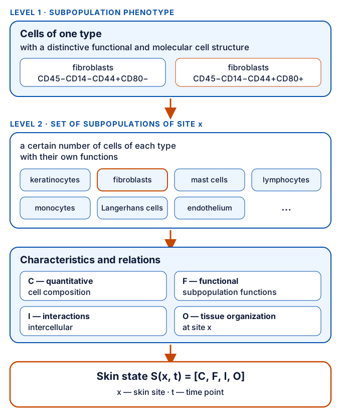
Diagram. Two levels of description and their relation to the skin state S(x, t). No numerical values.
What are the possibilities, limitations and open questions?
How phenotypes are studied. Subpopulation phenotypes are studied by flow cytometry in a cell suspension obtained from a skin biopsy specimen; the result is presented as a skin cytoimmunogram (in Russian, ЦИГК). A calculated indicator — the phenotypic index of skin inflammation (FIVK) — is built on individual phenotypes from this set.
Limitations.
- A phenotype is described by a limited set of markers. The author notes that the choice of a limited number of features “affects the accuracy of recognizing the diversity of cell features and, consequently, the diversity of cell functions.”
- The features make it possible to judge only individual aspects of the functional state of cells.
- The result refers to a skin fragment within the biopsy, not to the whole skin.
- Not all components of the conceptual model of the skin state are measured directly.
Open questions. The author formulates, for example, the following one: “What are the properties of cell phenotypes formed by combining certain proportions of cells of each type with their own functions?” This and other questions are collected on the page Open Research Questions.
Continue reading
- Skin State: From Cellular Phenotype to a Mathematical Model — what the skin state S(x, t) is, how it can be written down and how it differs from a diagnosis.
- The Skin Cytoimmunogram: From Cell Suspension to a Characteristic of the Skin State — how a skin cytoimmunogram is obtained, how the report form is structured and how to read its numbers.
- FIVK: Calculating and Interpreting the Inflammatory Component of the Skin State — how the index is calculated from 11 phenotypes and how its value should be understood.
See also: principles 1 and 2 · the concept · the terms “Skin cell subpopulation phenotype” and “Cell subpopulations” · overview of methods
Sources
Published sources
- Prototype of the website “Phenotypic Dermatology — a theory of skin states” (structure and wording of the sections; in Russian). — approved definition of the skin state (block “Key idea”) · Original
- Goltsov S. Fenotipicheskaya dermatologiya [Phenotypic Dermatology]. Tyumen: Publisher S. V. Goltsov, 2026. 208 p. ISBN 978-5-600-04919-2 (in Russian). — pp. 35, 42–43, 52–53, 55, 60 (principles 1 and 2), 66–70 (Example 1) · About the monograph · Original
- Goltsov S. V., Goltsova E. N., Sukhovey Yu. G., Kostolomova E. G., Pauls V. Yu. Cytoimmunogram of the skin: a new method for objective evaluation of the skin cell subpopulation profile [official English title of the journal article]. Medical Immunology (Russia). 2018; 20(3): 373–382. doi: 10.15789/1563-0625-2018-3-373-382 (article in Russian). — “Materials and methods” — detection of surface molecules and determination of the phenotype · Entry in Publications · Original
- Goltsov S. V., Kostolomova E. G., Sukhovey Yu. G., Pauls V. Yu. Sposob opredeleniya subpopulyatsionnogo sostava kletok kozhi i polucheniya tsitoimmunogrammy kozhi [A method for determining the subpopulation composition of skin cells and obtaining a skin cytoimmunogram]. Patent RU 2 630 607 C1. Application 2016121997 of 02.06.2016; registered and published 11.09.2017 (in Russian). — description and claims — the set of skin cytoimmunogram indicators · Patents · Original
The author’s clarifications and materials
- Goltsov S. V. Explanations of the notation S(x, t): a formal model of the skin state, notation of a transition, the role of the skin cytoimmunogram and FIVK, definition of diagnosis (author’s statement, PDF, 2026-11-18; in Russian). — the author’s explanations of 18 November 2026: S(x, t), the role of the skin cytoimmunogram
- Goltsov S. V. Scientific review of the article “What is the skin state S(x, t)” (author’s statement, 2026-11-18; in Russian). — the author’s review of 18 November 2026: “unit of analysis”, features and functions, the model [C, F, I, O]
- Goltsov S. V. The concept of Phenotypic Dermatology: text and brief for the concept diagram (author’s statement, 2026-11-18; in Russian). — text of the concept, paragraph 3
- Goltsov S. V. Agreed clarifications for the articles on the skin cell subpopulation phenotype, the skin cytoimmunogram and FIVK (author’s statements, 2026-11-20 and 2026-11-21; in Russian). — the author’s agreed clarifications of 20 November 2026: two levels of description; correspondence of the twelve cell types to the skin cytoimmunogram set; the rule for reading the “+” and “−” signs
This material is for information only and does not replace a consultation with a doctor.